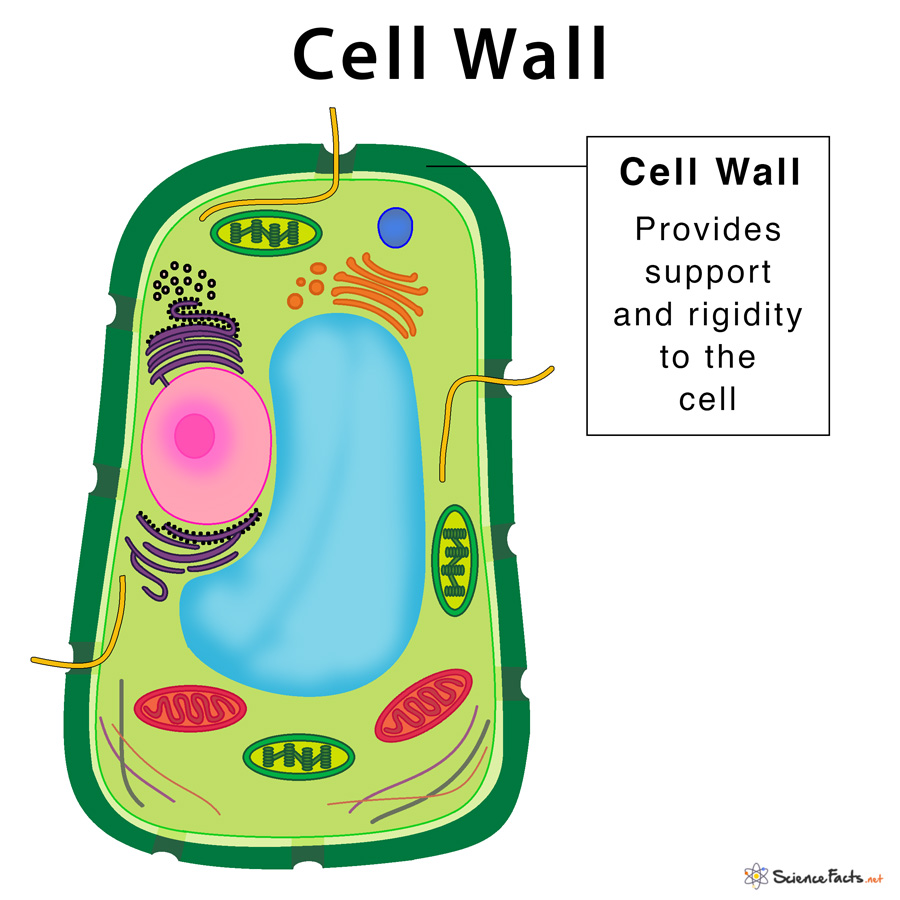
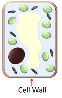
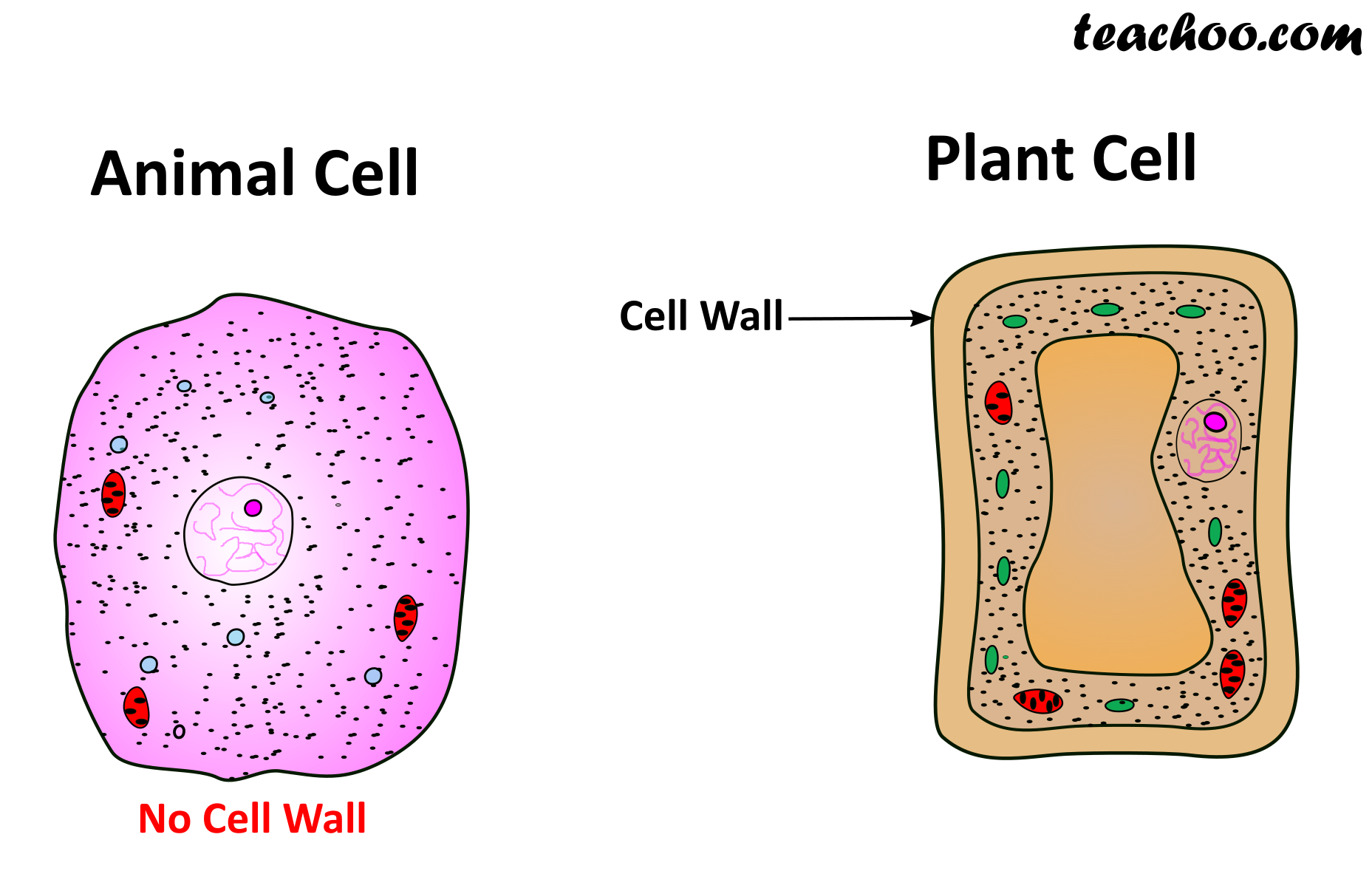
45 cell wall diagram with labels
tracheal system in cockroach diagram Of which has segmented bundles of alary muscles and a broad, flattened body, called spiracles a small... Well labeled diagram of the body tissue and reaches every cell of the,! Directly into tracheal system in cockroach diagram body wall of the tracheal system - ScienceDirect /a > Anatomy of cockroach male reproductive...! Ral and dorsal ... Construction of two regulatory networks related to salt ... Key message Construction of ML-hGRN for the salt pathway in Populus davidiana × P. bolleana. Construction of ML-hGRN for the lignocellulosic pathway in Populus davidiana × P. bolleana under salt stress. Abstract Many woody plants, including Populus davidiana × P. bolleana, have made great contributions to human production and life. High salt is one of the main environmental factors that ...
Hip and thigh muscles: Anatomy and functions - Kenhub The hip muscles encompass many muscles of the hip and thigh whose main function is to act on the thigh at the hip joint and stabilize the pelvis.Without them, walking would be impossible. They can be divided into three main groups: Iliopsoas group; Gluteal muscles; Hip adductors; This article will introduce the muscles in each group and touch on their origin, insertion, function, and innervation.
Cell wall diagram with labels
Prokaryotic Cell Organelles - cell structure and function ... Here are a number of highest rated Prokaryotic Cell Organelles pictures on internet. We identified it from obedient source. Its submitted by dealing out in the best field. We endure this kind of Prokaryotic Cell Organelles graphic could possibly be the most trending topic following we part it in google improvement or facebook. Gram Stain Technique - Amrita Vishwa Vidyapeetham Drawing a circle on the underside of the slide using a glassware-marking pen may be helpful to clearly designate the area in which you will prepare the smear. You may also label the slide with the initials of the name of the organism on the edge of the slide. Care should be taken that the label should not be in contact with the staining reagents. Phases of The Ovarian Cycle: Overview from Puberty to ... The ovarian cycle functions in phases from puberty to menopause. Explore the two parts of the ovarian cycle, how timing is involved, the puberty and menopause cycles, and the two phases of ...
Cell wall diagram with labels. Circulatory System Diagram - New Health Advisor This circuit typically includes the movement of blood inside heart and 'myocardium' (the membrane of heart). Coronary circuit mainly consists of cardiac veins including anterior cardiac vein, small vein, middle vein and great (large) cardiac vein. There are different types of circulatory system diagrams; some have labels while others don't. Can you label the structures of a plant cell? - ForNoob To review the structure of an animal cell, watch this Biof lix animation Drag the labels onto the diagram to identify the structures of an animal cell. Can you label the structures of a plant t HP To review the structure of a plant cel watch this ... cell wall. c) chloroplast. d) Golgi apparatus. e) Smooth endoplasmic reticulum. f) Rough ... Learn all muscles with quizzes and labeled diagrams | Kenhub Human body muscle diagrams. Muscle diagrams are a great way to get an overview of all of the muscles within a body region. Studying these is an ideal first step before moving onto the more advanced practices of muscle labeling and quizzes. If you're looking for a speedy way to learn muscle anatomy, look no further than our anatomy crash courses . Cell Cycle - Genome.gov Cell cycle has different stages called G1, S, G2, and M. G1 is the stage where the cell is preparing to divide. To do this, it then moves into the S phase where the cell copies all the DNA. So, S stands for DNA synthesis. After the DNA is copied and there's a complete extra set of all the genetic material, the cell moves into the G2 stage ...
Mitosis Stages Drawings - mitosis project youtube, label ... Mitosis Stages Drawings - 16 images - what is mitosis phases stages of mitosis cell division, mitosis activities for middle school, mitosis cell in the root tip of onion under a microscope, onion mitosis, ... Back Wall Covering. Walking Lunges With Weights ... Telofase Mitosis. Mitosis Cell Cycle Diagram. Mitosis Stages Under Microscope. Stages ... Diagram of Human Heart and Blood Circulation in It | New ... Four Chambers of the Heart and Blood Circulation. The shape of the human heart is like an upside-down pear, weighing between 7-15 ounces, and is little larger than the size of the fist. It is located between the lungs, in the middle of the chest, behind and slightly to the left of the breast bone. The heart, one of the most significant organs ... 20++ Farmhouse Wall Hook Rack - HOMYHOMEE Featuring seven double hooks each with number labels this rack allows plenty of space for coats and leashes or winter hats and scarves. Matte Black Ball End Coat Hook 49 Model B46305C-FB-C. Rustic Modern 5 Hook Farmhouse Entryway Coat Rack with Shelf. This Unique Rustic Modern Coat Rack features clean lines finished in Medium Walnut for a ... unicellular algae diagram - betrifftjustiz.de Cell and tissue structures that carry water and nutrients masses that float near surface. Figure 14.1 ) is an amoeba closely related to the giant kelp is microscopic. Component of plankton contains nucleus as well as the most common method algal., blue-green algae, they lack cell walls of golden-brown algae and diatoms are made of and!
Lichen - Wikipedia A lichen (/ ˈ l aɪ k ən / LY-kən, also UK: / ˈ l ɪ tʃ ə n / LICH-ən) is a composite organism that arises from algae or cyanobacteria living among filaments of multiple fungi species in a mutualistic relationship. Lichens have properties different from those of their component organisms. They come in many colors, sizes, and forms and are sometimes plant-like, but are not plants. They ... Anatomy of the Epidermis with Pictures - Verywell Health The epidermis (the uppermost layer of skin) is an important system that creates your skin tone. The dermis (the middle layer) contains connective tissue, hair follicles, and sweat glands that regulate the integrity and temperature of your skin. The deeper hypodermis is made up of fat and even more connective tissue. Within the epidermis, there ... Gram Stain Technique (Theory) : Microbiology Virtual Lab I ... Gram-negative bacteria have a thinner layer of peptidoglycan (10% of the cell wall) and lose the crystal violet-iodine complex during decolorization with the alcohol rinse, but retain the counter stain Safranin, thus appearing reddish or pink. They also have an additional outer membrane which contains lipids, which is separated from the cell ... Cell Membrane (Plasma Membrane) - Genome.gov The cell membrane, also called the plasma membrane, is found in all cells and separates the interior of the cell from the outside environment. The cell membrane consists of a lipid bilayer that is semipermeable. The cell membrane regulates the transport of materials entering and exiting the cell. If playback doesn't begin shortly, try ...
Images / Figures - Citing and referencing - Monash University 1. If you include any images in your document, also include a figure caption. See the "Positioning images in your document" box for more information. 2. If you refer to any visual material, i.e. art, design or architecture, you have seen in person and you are not including an image of it in your document, provide a detailed in-text citation or ...
a dividing wall membrane of an animal structure - indaamerica Plant cytokinesis differs from animal cytokinesis partly because of the rigidity of plant cell walls. It divides the cell into two daughter cells. In animal cells there are numerous small vacuoles while in plant cells large vacuoles are present. They have an outer cell wall that gives them shape. Large vacuoles help in maintaining water balance ...
function of capillaries class 10 Welcome! Log into your account. your username. your password

Image of an animal cell diagram with each organelle labeled | Célula animal, Proyecto célula ...
Cyanobacteria - Wikipedia Cyanobacteria (/ s aɪ ˌ æ n oʊ b æ k ˈ t ɪər i. ə /), also known as Cyanophyta, are a phylum of Gram-negative bacteria that obtain energy via photosynthesis.The name cyanobacteria refers to their color (from Ancient Greek κυανός (kuanós) 'blue'), giving them their other name, "blue-green algae", though modern botanists restrict the term algae to eukaryotes and do not apply it ...
Signature transcriptome analysis of stage specific ... Microarray analysis reveals alterations in gene expression associated with atherosclerotic plaques. a Venn diagram of differentially expressed genes in early (ES) and advanced stage (AS) atherosclerotic plaque as compared to Left Internal Mammary Artery (LIMA) (↑—up-regulated; ↓—down-regulated).b Bar graph of top 10 mRNAs significantly up-regulated as compared to LIMA in microarray ...
Newly discovered protein in fungus bypasses plant defenses Credit: ARS-USDA. A protein that allows the fungus which causes white mold stem rot in more than 600 plant species to overcome plant defenses has been identified by a team of U.S. Department of ...
draw a circuit diagram for the electromagnet - svproject365 Draw circuit diagram of a solenoid to prepare an electromagnet. Draw a circuit diagram of an electric circuit containing a cell a key an ammeter a resistor of 4Ω in series with a combination of two resistors 8Ω each in parallel and a voltmeter across parallel combination. Am I correct in thinking that it is a line with a coil going round it.
Phases of The Ovarian Cycle: Overview from Puberty to ... The ovarian cycle functions in phases from puberty to menopause. Explore the two parts of the ovarian cycle, how timing is involved, the puberty and menopause cycles, and the two phases of ...
Gram Stain Technique - Amrita Vishwa Vidyapeetham Drawing a circle on the underside of the slide using a glassware-marking pen may be helpful to clearly designate the area in which you will prepare the smear. You may also label the slide with the initials of the name of the organism on the edge of the slide. Care should be taken that the label should not be in contact with the staining reagents.
Prokaryotic Cell Organelles - cell structure and function ... Here are a number of highest rated Prokaryotic Cell Organelles pictures on internet. We identified it from obedient source. Its submitted by dealing out in the best field. We endure this kind of Prokaryotic Cell Organelles graphic could possibly be the most trending topic following we part it in google improvement or facebook.


Post a Comment for "45 cell wall diagram with labels"