42 human heart diagram and labels
A Diagram of the Heart and Its Functioning Explained in ... The heart blood flow diagram (flowchart) given below will help you to understand the pathway of blood through the heart.Initial five points denotes impure or deoxygenated blood and the last five points denotes pure or oxygenated blood. 1.Different Parts of the Body ↓ 2.Major Veins ↓ 3.Right Atrium ↓ 4.Right Ventricle ↓ 5.Pulmonary Artery ↓ 6.Lungs File:Diagram of the human heart.svg - Wikimedia Commons Description. Diagram of the human heart.svg. SVG illustration of the human heart, created by Wapcaplet in Sodipodi. Slightly modified for correct rendering by Yaddah (no changes to content). Cropped version withour white space available at File:Diagram of the human heart (cropped).svg Uploaded on 24 Dec 2003.
13+ Heart Diagram Templates - Sample, Example, Format ... Free heart diagrams can be helpful for students in understanding the heart and its functioning. Human heart is a complicated figure and for students from science, they will often need the images of the heart for its illustration. The above collection of heart samples will make it easier for students to download, print and use it in their projects.

Human heart diagram and labels
Heart Diagram - 15+ Free Printable Word, Excel, EPS, PSD ... Heart Diagram - 15+ Free Printable Word, Excel, EPS, PSD Template Download A heart diagram is a popular design used by different people for various uses. It can be used by a teacher or student for academic purpose, by a friend or relative for mutually sending and exchanging cards or for baby toys or printing on dresses etc. Label the heart - Science Learning Hub In this interactive, you can label parts of the human heart. Drag and drop the text labels onto the boxes next to the diagram. Selecting or hovering over a box will highlight each area in the diagram. Pulmonary vein Right atrium Semilunar valve Left ventricle Vena cava Right ventricle Pulmonary artery Aorta Left atrium Download Exercise Tweet Simple Diagram of the Heart Labelling Activity - Twinkl This simple heart diagram with labels is a fab learning activity to help your pupils aged 10-11 understand the heart and its function in the human body.
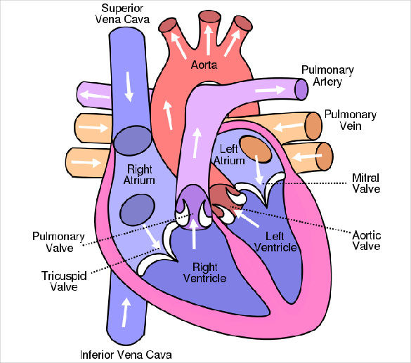
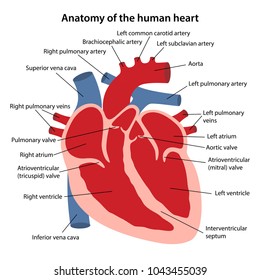
Human heart diagram and labels. Human Heart Diagram Class 10 | Get Easy Tricks to Draw ... Human Heart Diagram with Label In your exams, your diagram will be marked only if you have labeled it. That is how important labels are. If you practice the image multiple times with the labels, you will automatically remember all the marking labels. And to add to it, you must make sure all your marking labels are aligned. The Human Heart Cardiovascular System Labeling Worksheet Using the blank heart diagram students are asked to label the aorta, superior vena cava, pulmonary arteries, pulmonary veins, atrium, ventricles, and aortic valves. This simple human heart diagram could be used as both a starter or plenary in order to assess students prior and post knowledge of the structure of the heart. Human Heart Diagram Without Labels - Labelling Worksheet The human heart is a muscle made up of four chambers, these are: Two upper chambers - the left atrium and right atrium Two lower chambers - the left and right ventricles. It's also made up of four valves - these are known as the tricuspid, pulmonary, mitral and aortic valves. Heart Diagram with Labels and Detailed Explanation Heart Detailed Diagram Heart - Chambers There are four chambers of the heart . The upper two chambers are the auricles and the lower two are called ventricles. The two atria are thin-walled chambers that receive blood from the veins. The two ventricles are in contrast thick-walled which forcefully pump blood out of the heart.
File:Diagram of the human heart (cropped).svg - Wikipedia File:Diagram of the human heart (cropped).svg. Size of this PNG preview of this SVG file: 611 × 600 pixels. Other resolutions: 244 × 240 pixels | 489 × 480 pixels | 782 × 768 pixels | 1,043 × 1,024 pixels | 2,086 × 2,048 pixels | 663 × 651 pixels. This is a file from the Wikimedia Commons. Information from its description page there is ... Human Heart Diagram Pictures, Images and Stock Photos Human Heart Diagram Pictures, Images and Stock Photos View human heart diagram videos Browse 3,849 human heart diagram stock photos and images available, or search for heart illustration or pulmonary artery to find more great stock photos and pictures. Newest results heart illustration pulmonary artery kidney diagram Human Heart Circulatory System New * The Human Heart Labelling Worksheet - Twinkl There are two versions of this resource. The standard version comes with two pages - the first has a human heart diagram without labels, and the second has the ... Diagram of Human Heart and Blood Circulation in It | New ... Exterior of the Human Heart A heart diagram labeled will provide plenty of information about the structure of your heart, including the wall of your heart. The wall of the heart has three different layers, such as the Myocardium, the Epicardium, and the Endocardium. Here's more about these three layers. Epicardium
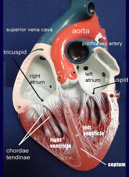
PDF Anatomy of Heart Labeled and Unlabeled Images Anatomy of Heart Labeled and Unlabeled Images 2 3 4 5 6 7 8 9 10 11 Mediastinum Superior vena cava Ribs Right lung Diaphragm Right and left atrioventricular sulci (a) Location of heart in chest, anterior view © 2019 Pearson Education, Inc Aorta Pulmonary trunk Retractor Anterior interventricular sulcus Heart Structure | BioNinja Recognition of the chambers and valves of the heart and the blood vessels connected to it in dissected hearts or in diagrams of heart structure. File:Diagram of the human heart (no labels).svg ... File:Diagram of the human heart (no labels).svg. Size of this PNG preview of this SVG file: 498 × 599 pixels. Other resolutions: 199 × 240 pixels | 399 × 480 pixels | 499 × 600 pixels | 639 × 768 pixels | 851 × 1,024 pixels | 1,703 × 2,048 pixels | 533 × 641 pixels. A Labeled Diagram of the Human Heart You Really Need to ... The human heart, comprises four chambers: right atrium, left atrium, right ventricle and left ventricle. The two upper chambers are called the left and the right atria, and the two lower chambers are known as the left and the right ventricles. The two atria and ventricles are separated from each other by a muscle wall called 'septum'.
File:Heart diagram-en.svg - Wikipedia File:Heart diagram-en.svg. Size of this PNG preview of this SVG file: 762 × 600 pixels. Other resolutions: 305 × 240 pixels | 610 × 480 pixels | 976 × 768 pixels | 1,280 × 1,008 pixels | 2,560 × 2,015 pixels | 893 × 703 pixels. This is a file from the Wikimedia Commons. Information from its description page there is shown below.
Heart Labeling Quiz: How Much You Know About Heart ... Create your own Quiz Here is a Heart labeling quiz for you. The human heart is a vital organ for every human. The more healthy your heart is, the longer the chances you have of surviving, so you better take care of it. Take the following quiz to know how much you know about your heart. Questions and Answers 1. What is #1? 2. What is #2? 3.
Human Heart: Label the diagram 1 worksheet Human Heart: Label the diagram 1 Study the figure carefully.Label the 10 parts of the human heart A-J.
Human Heart-Label Diagram | Quizlet Label the diagram Learn with flashcards, games, and more — for free.
Human Heart Diagram Without Labels | Human heart diagram ... Human Heart Diagram Without Labels. Find this Pin and more on AnP by Susan Wells. This is Page 39 of a photographic atlas I created as a laboratory study resource for my BIOL 121 Anatomy and Physiology I students on the bones and bony landmarks of the axial skeleton. Credits: All photography, text, and labels by Rob Swatski, Assistant Professor ...
Human Heart Diagram Photos and Premium High Res Pictures ... Browse 612 human heart diagram stock photos and images available, or search for heart illustration or pulmonary artery to find more great stock photos and pictures. An anatomical diagram showing the arteries of the human heart, circa 1930. Diagram of the human brain and other organs.
How to Draw a Human Heart: 11 Steps (with Pictures) - wikiHow The heart works like a pump and beats 100,000 times a day. The heart has two sides, separated by an inner wall called the septum. The right side of the heart pumps blood to the lungs to pick up oxygen. The left side of the heart receives the oxygen-rich blood from the lungs and pumps it to the body.
Human Heart Diagram - Human Body Pictures - Science for Kids Photo description: This is an excellent human heart diagram which uses different colors to show different parts and also labels a number of important heart component such as the aorta, pulmonary artery, pulmonary vein, left atrium, right atrium, left ventricle, right ventricle, inferior vena cava and superior vena cava among others.
Human Heart - Diagram and Anatomy of the Heart The heart is a muscular organ about the size of a closed fist that functions as the body's circulatory pump. It takes in deoxygenated blood through the veins and delivers it to the lungs for oxygenation before pumping it into the various arteries (which provide oxygen and nutrients to body tissues by transporting the blood throughout the body).
Human Heart Diagram Labeled | Science Trends Human Heart Diagram Labeled Daniel Nelson 1, January 2019 | Last Updated: 3, March 2020 The human heart is an organ responsible for pumping blood through the body, moving the blood (which carries valuable oxygen) to all the tissues in the body. Without the heart, the tissues couldn't get the oxygen they need and would die.
Heart Diagram with Labels and Detailed Explanation The human heart is the most crucial organ of the human body. It pumps blood from the heart to different parts of the body and back to the heart. The most common heart attack symptoms or warning signs are chest pain, breathlessness, nausea, sweating etc. The diagram of heart is beneficial for Class 10 and 12 and is frequently asked in the ...
Heart Anatomy: Labeled Diagram, Structures, Blood Flow ... There are 4 chambers, labeled 1-4 on the diagram below. To help simplify things, we can convert the heart into a square. We will then divide that square into 4 different boxes which will represent the 4 chambers of the heart. The boxes are numbered to correlate with the labeled chambers on the cartoon diagram.
PDF Free Anatomy Coloring Page - NCSU Color Me. The ate.2S the heart With oxygen ate labeled with at'l Color these areas The areas o' the heart with less oxygen ate labeled with a color areas BLUE. ARTERY LEFT LUNG PULMONARY VEINS AORTA PULMONARY VEINS raGHT LUNG ATRIUM RIGHT VENTRICLE INFERIOR VFNACAVA LEFT LEFT VENTRICLE AORTA BODY Downloaded from azcoloring.com
Human Heart (Anatomy): Diagram, Function, Chambers ... The heart is a muscular organ about the size of a fist, located just behind and slightly left of the breastbone. The heart pumps blood through the network of arteries and veins called the...

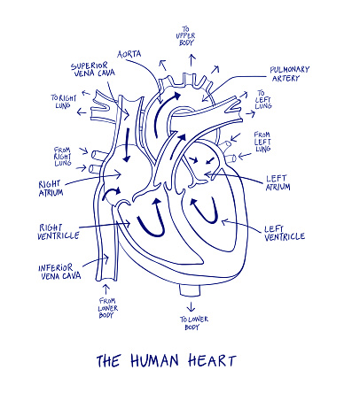
Human Heart Anatomy Diagram Blue Line On A White Background Stock Illustration - Download Image ...
Simple Diagram of the Heart Labelling Activity - Twinkl This simple heart diagram with labels is a fab learning activity to help your pupils aged 10-11 understand the heart and its function in the human body.
Label the heart - Science Learning Hub In this interactive, you can label parts of the human heart. Drag and drop the text labels onto the boxes next to the diagram. Selecting or hovering over a box will highlight each area in the diagram. Pulmonary vein Right atrium Semilunar valve Left ventricle Vena cava Right ventricle Pulmonary artery Aorta Left atrium Download Exercise Tweet
Heart Diagram - 15+ Free Printable Word, Excel, EPS, PSD ... Heart Diagram - 15+ Free Printable Word, Excel, EPS, PSD Template Download A heart diagram is a popular design used by different people for various uses. It can be used by a teacher or student for academic purpose, by a friend or relative for mutually sending and exchanging cards or for baby toys or printing on dresses etc.
![BIOMED ALL INVITED: The Human Heart [ANATOMY/PHYSIOLOGY/CONDUCTION SYSTEM]](https://blogger.googleusercontent.com/img/b/R29vZ2xl/AVvXsEjQgi4mVEqqHg0V3SLwXc_SNLTR5VD5JdiMOD_BaEl2E1fzFNjSD-B3AJggoZ5VjWJD4CQKi5EXpMMvJE8e9ryZfrOvysVpTjTNG9DFc2JUO6XMMDNUH_7aEGDdXUyDWYqvvSMvimroBW9h/s1600/heart+anatomy.jpg)


Post a Comment for "42 human heart diagram and labels"