41 spinal cord model with labels
Labeled Brain Model Diagram | Science Trends The flocculonodular lobe Is the oldest part of the cerebellum in terms of evolution , and this is the structure responsible for spatial orientation and balance. The medial region of the posterior and anterior lobes function to control fine body movements, taking in input from the spinal cord as well as the auditory and visual systems of the brain. Spinal cord transverse section coverings label 3D model - CGTrader Spinal cord transverse section coverings label 3D model, available formats , ready for 3D animation and other 3D projects | CGTrader.com ... A blend model of spinal cord along with it covering layers and nerve roots. The ascending and descending tracts of spinal cord transverse section are labelled in detail. The material is image textures with ...
Spinal Cord Model - YouTube Dixon discusses Enlargements, cord, pia, dura, mater, denticulate ligaments, 8 spinal nerves, cauda equina, sympathetic chain ganglion , paravetrebral gangli...

Spinal cord model with labels
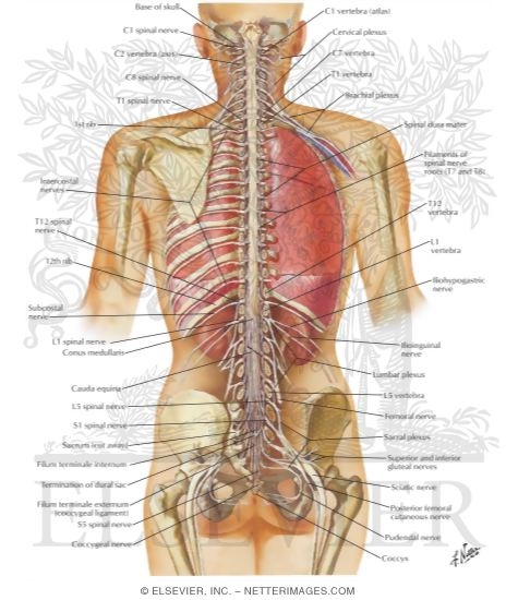
Spinal Cord Labeling Quiz - PurposeGames.com This is an online quiz called Spinal Cord Labeling. There is a printable worksheet available for download here so you can take the quiz with pen and paper. Your Skills & Rank. Total Points. 0. Get started! Today's Rank--0. Today 's Points. One of us! Game Points. 12. You need to get 100% to score the 12 points available. Spinal Cord - Anatomy, Structure, Function, & Diagram In adults, the spinal cord is usually 40cm long and 2cm wide. It forms a vital link between the brain and the body. The spinal cord is divided into five different parts. Several spinal nerves emerge out of each segment of the spinal cord. There are 8 pairs of cervical, 5 lumbar, 12 thoracics, 5 sacral and 1 coccygeal pair of spinal nerves. Spinal Cord in the Spinal Canal (BS 31) · Anatomy models | SOMSO® Spinal Cord in the Spinal Canal. Seen from the ventral side, natural size, in SOMSO-PLAST®. The model shows the brain stem and the spinal cord, as well as the nerve branches, up to the coccygeal plexus. On the left side, the sympathetic trunk with its connections to the central nervous system is shown. In one piece.
Spinal cord model with labels. Spinal Cord Models - San Diego Mesa College Spinal Cord Models. Click on a photo for a larger view of the model. Click on Label for the labeled model. Back to Nervous System. Spinal Cord (transverse section) Spinal Cord (close up) Spinal Cord (longitudinal view) Label: Label: Label: Spinal Cord (superior ls) Spinal Cord (inferior ls) › jdownloads › GET_TaskGRADE: 5 SUBJECT: NATURAL SCIENCES AND TECHNOLOGY TERM ONE ... o Backbone - protects the spinal cord o Ribs - protects the lungs and heart o Shoulder blade, legs and hips - allow for movement . Key concepts you have observed about structures: Shell structures are formed from one piece of material. Frame structures are made from different parts that are joined together. spinal cord anatomy, labeling spinal model Quiz This is an online quiz called spinal cord anatomy, labeling spinal model. There is a printable worksheet available for download here so you can take the quiz with pen and paper. Your Skills & Rank. Total Points. 0. ... label eye muscles and structures 10p Image Quiz. internal ear anatomy 15p Image Quiz. spinal nerves and plexuses 11p Image Quiz. PDF Spinal Cord Classroom Teacher - Duquesne University identify different parts of the spinal cord by building their own spinal columns out of string and empty spools of thread. In addition, students will label the parts of the spinal cord. Time 35 minutes Activity Summary: Spinal Column Concentration Students will review spinal cord key terms and work in pairs to play the Spinal Cord Concentration ...
Spinal Cord Diagram with Detailed Illustrations and Clear Labels The spinal cord is one of the most important structures in the human body. In fact, it is the most important structure for any vertebrates. Anatomically, the spinal cord is made up is made up of nervous tissue and is integrated into the spinal column of the backbone. Main Article: Spinal Cord - Anatomy, Structure, Function, and Spinal Cord Nerves PDF Anatomy & Physiology - TMCC Somso Model QS 61 Wedge-shaped segment from the compact part of a long bone 1. Periosteum 2. Outer general lamellae 3. Perforating fibers of Sharpey spinal cord lab model Diagram | Quizlet Start studying spinal cord lab model. Learn vocabulary, terms, and more with flashcards, games, and other study tools. › service-details › frequently-askedFrequently asked questions about car seats | Mass.gov The seat has labels stating date of manufacture and model number. You need this information to find out if there is a recall on the car seat or if the seat has expired. The seat has no recalls. If you do find a recall on the car seat, you should contact the manufacturer. The seat has all its parts.
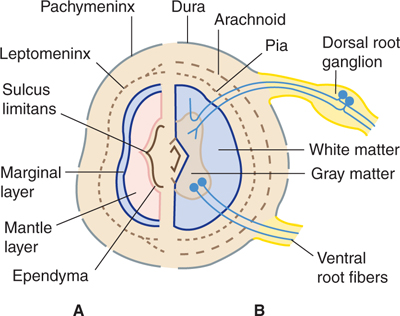
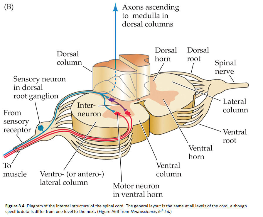
Spinal cord transverse section coverings label 3D Model Model's Description: Spinal cord transverse section coverings label 3d model contains 2,035,824 polygons and 835,146 vertices. Please wait for full texture to load (HD). A blend model of spinal cord along with it covering layers and nerve roots. The ascending and descending tracts of spinal cord transverse section are labelled in detail. Nervous System Models | Spinal Cord with Nerves Models Deluxe Spinal Cord Model (0165-00) Item # DGA65. $369.00 $339.00. Add to cart. Kyoto Kagaku Full-Figure Nervous System Model. Item # KK-A25. Add to cart. Kyoto Kagaku Nerves and Vessels of Arm Model. Item # KK-A144. Add to cart. Physiology of Nerves Series, 5 magnetics - illustrated metal board ... PDF Anatomy and Physiology of the Spinal Cord Spinal Cord Injury - Traumatic Causes Australia 2008-2009 The Spinal Column Your vertebrae (neck and back bones) form a circular column to protect the spinal cord. There are 33 vertebrae, starting at the base of the skull, and ending with two sections of joined/fused vertebrae Anatomy of the Spinal Cord (Section 2, Chapter 3) Neuroscience Online ... The spinal cord extends from the foramen magnum where it is continuous with the medulla to the level of the first or second lumbar vertebrae. It is a vital link between the brain and the body, and from the body to the brain. The spinal cord is 40 to 50 cm long and 1 cm to 1.5 cm in diameter. Two consecutive rows of nerve roots emerge on each of ...
opentext.wsu.edu › psych105nusbaum › chapter90 Diagnosing and Classifying Psychological Disorders THE DIAGNOSTIC AND STATISTICAL MANUAL OF MENTAL DISORDERS (DSM). Although a number of classification systems have been developed over time, the one that is used by most mental health professionals in the United States is the Diagnostic and Statistical Manual of Mental Disorders (DSM-5), published by the American Psychiatric Association (2013).

Spinal Cord Model: Dura Mater, Arachnoid Mater, Pia Mater, Ventral ... | Histology - Spinal Cord ...
SPINAL CORD MODEL Flashcards | Quizlet Objectives for Spinal Cord (fifth cervic…. 210: Chapter 11 Blended Skills and Critical Thinki…. 5th - Social Studies Review - ch7 notes and questi….
› en › e-AnatomyArm, forearm, and hand: MRI of anatomy - e-Anatomy - IMAIOS Sep 13, 2021 · By moving the mouse cursor over a particular area of the arm or forearm, this area is highlighted and the labels are displayed: anterior, lateral or posterior compartment. On the vertical left menu, a medical illustration of an upper limb skeleton based on a three dimensional (3D) model simplifies the access to the anatomical regions.
› inflammation-autoimmunityIs Chronic Fatigue Syndrome Autoimmune? Inflammatory? Dec 04, 2020 · In molecular mimicry, the immune system fights an infectious agent and then begins to confuse it with a similar cell in the body and begins attacking it. Essentially, because both cells look similar, the immune system labels them as identical, when in fact one type actually belongs in your body.
Spinal cord: Anatomy, structure, tracts and function | Kenhub Anatomy. The spinal cord is part of the central nervous system (CNS). It is situated inside the vertebral canal of the vertebral column. During development, there's a disproportion between spinal cord growth and vertebral column growth. The spinal cord finishes growing at the age of 4, while the vertebral column finishes growing at age 14-18.
Labeled Spinal Cord Model at Anatomy The spinal cord is 40 to 50 cm long and 1 cm to 1.5 cm in diameter. Several spinal nerves emerge out of each segment of the spinal cord. Source: faculty.etsu.edu. Anatomy of the spinal cord it connects the brain and spinal cord to the skull and spinal canal. Two consecutive rows of nerve roots emerge on each of.
Spinal Cord Quiz: Cross-Sectional Anatomy - GetBodySmart Spinal Cord - Cross-Sectional Anatomy. Start Quiz. Want to learn faster? Look no further than these interactive, exam-style anatomy quizzes. Learn anatomy faster and remember everything you learn. Start Now. Related Articles. Parts of the Brain Quiz. Test your knowledge with the parts of the brain and their functions in a fun and interactive ...
Spinal cord | Encyclopedia | Anatomy.app | Learn anatomy | 3D models ... Conus medullaris, cauda equina and filum terminale. The spinal cord is shorter than the vertebral column and it ends at the first or second lumbar vertebrae (L1/L2) level as conus medullaris (medullary cone). The conus medullaris is a cone-shaped termination of the spinal cord that is connected to the coccyx by a fibrous connective tissue strand called filum terminale.
Spinal cord - Wikipedia The spinal cord is a long, thin, tubular structure made up of nervous tissue, which extends from the medulla oblongata in the brainstem to the lumbar region of the vertebral column.It encloses the central canal of the spinal cord, which contains cerebrospinal fluid.The brain and spinal cord together make up the central nervous system (CNS). In humans, the spinal cord begins at the occipital ...
Axis Scientific Spine Model, 34" Life Size Spinal Cord Model with ... Axis Scientific Spine Model, 34" Life Size Spinal Cord Model with Vertebrae, Nerves, Arteries, Lumbar Column, and Male Pelvis, Includes Stand, Detailed Product Manual and Worry Free 3 Year Warranty ... Full Color Spine Model Study Guide . Includes 26 labeled parts! Read more. Read more. Read more. Videos. Page 1 of 1 Start Over Page 1 of 1 ...
› topics › neuroscienceGliosis - an overview | ScienceDirect Topics H. Richard Winn MD, in Youmans and Winn Neurological Surgery, 2017. Inflammatory Mechanisms and Gliosis. TBI produces neuroinflammation in the CNS in which rupture of the blood-brain barrier leads to accumulation of leukocytes from the systemic circulation and subsequent initiation of the immune functions of native glia. 20 Acute inflammatory cytokine-mediated events such as monocyte ...
Spinal Cord Anatomy Model Labeled at Anatomy Spinal Cord Anatomy Model Labeled. Learn spinal cord model brain anatomy with free interactive flashcards. The spinal cord is a long, thin, tubular structure made up of nervous tissue, which extends from the medulla oblongata in the brainstem to the lumbar region of the vertebral column.
Spinal Cord | Trunk Wall (Part 1) | Vertebral Column | Bones of the ... The spinal cord models are 5-times life-size. Orient yourself when looking at the spinal cord, the cross-sectional images will be on the right, the 3B symbol will be on the top left of the base. While looking at the spinal cord model in this position, the side closest to you is ventral, the side furthest away from you is dorsal. ...
9,901 Spinal Cord Stock Photos and Images - 123RF Download spinal cord stock photos. Affordable and search from millions of royalty free images, photos and vectors. ... Model of a human spine, spinal columns. X-ray C-SPINES : AP, LATERAL showing S/p internal fixation C4, C5 & C6 with plate & screws. ... Back anatomy labeled. Human spinal nerves indicated with white points on female torso ...
Learn the spinal cord with diagrams and quizzes | Kenhub The spinal cord, along with the brain, makes up the central nervous system (CNS). It is a long tubular structure comprised of nervous tissue, extending from the cervical to the lumbar region of the vertebral column. Just like other parts of the CNS, the spinal cord is comprised of white and gray matter. Spinal cord gray matter is the central ...


Post a Comment for "41 spinal cord model with labels"