38 images of compound microscope with labels
Compound Microscope Stock Photos and Images - Alamy Find the perfect compound microscope stock photo. Huge collection, amazing choice, 100+ million high quality, affordable RF and RM images. ... Compound Microscope Stock Photos and Images (1,984) compound microscope isolated. Related searches: ... Method of illuminating compound microscope with gas lamp. Labels: C, ... Compound Microscope Parts - Labeled Diagram and their Functions Basically, compound microscopes generate magnified images through an aligned pair of the objective lens and the ocular lens. In contrast, "simple microscopes" have only one convex lens and function more like glass magnifiers. [In this figure] Two "antique" microscopes played significant roles in the history of biology.
› publication › 320945390(PDF) Introduction to Microscopy - ResearchGate Nov 08, 2017 · • In compound microscope it will be i.e 10 X, f= 16 mm; 40 X, f= 4 mm; 100 X, f= 1.8 mm. • Image produced by objective lens falls on the eyepiece lens serve as objec t. • Image formed in the ...

Images of compound microscope with labels
Microscope Images at Various Magnifications | Microscope World Resources The different images below were taken with two different types of microscopes. The images of Paulownia wood, hair, and frog's blood were captured with a high power compound microscope using a Nikon camera adapter. The compound microscope typically has three or four magnifications - 40x, 100x, 400x, and sometimes 1000x. Compound Microscope Labeled Diagram | Quizlet QUESTION. The total magnification of a specimen being viewed with a 10X ocular lens and a 40X objective lens is. 15 answers. QUESTION. a mosquito beats its wings up and down 600 times per second, which you hear as a very annoying 600 Hz sound. if the air outside is 20 C, how far would a sound wave travel between wing beats. 2 answers. Parts of a Compound Microscope - Labeled (with diagrams) A compound microscope is known as a high-power microscope that enables you to achieve a high level of magnification. Smaller specimens can be thoroughly viewed using a compound microscope. ... Image 3: A compound microscope with a corresponding label of the different parts. imagesource: images.slideplayer.com ... Labels: microsopes Newer Post ...
Images of compound microscope with labels. Parts of a Compound Microscope and Their Functions - NotesHippo The main parts of compound microscope are the condenser lens, the objective lens, and the eyepiece lens, and these instruments are referred to as compound microscopes. Each of these components is made up of microscope lens combinations that are required to produce magnified images with minimal artefacts and aberrations. Structure of Microscope Simple Microscope - Parts, Functions, Diagram and Labelling Compound microscope - It comes with more than one lens and provides better magnification than the simple microscope. A compound microscope is also called a bright field microscope. It can provide magnification by up to 1,000 times. Stereo microscope/dissecting microscope - It can magnify objects by up to 300 times. It is used to visualize ... Solved Label the image of a compound light microscope using - Chegg Experts are tested by Chegg as specialists in their subject area. We review their content and use your feedback to keep the quality high. Transcribed image text: Label the image of a compound light microscope using the terms provided. Amazing 27 Things Under The Microscope With Diagrams - Microbe Notes The tail is transparent and thus is difficult to detect under a low-power microscope. 23. Spirogyra under the microscope. Spirogyra is a green alga found mostly in freshwater in the form of green clumps. Spirogyra is unicellular, but because it clumps together, it can be seen in the pond even with our naked eyes.
Place the labels on the image of a compound light | Chegg.com Expert Answer. Transcribed image text: Place the labels on the image of a compound light microscope. Group 1 labels identify the parts; group 2 labels identify the functions. Coaxial stage controls Stage clip Secures the slide on the stage Move the slide night eft and backwardforward Group 1 Group 2 Group 1 Group 2 Reset Help. Previous question. › seterra › en-anMicroscope Components - Science Quiz - GeoGuessr Microscope Components - Science Quiz: The most common type of modern microscope is called a compound microscope. They have two systems of lenses, one is the eyepiece and the other is comprised of one or more objective lenses. This type of microscope has become so advanced that some are capable of magnifying up to 1000 times! Microscopes are used in almost all types of scientific research, and ... Compound Microscope- Definition, Labeled Diagram, Principle, Parts, Uses In order to ascertain the total magnification when viewing an image with a compound light microscope, take the power of the objective lens which is at 4x, 10x or 40x and multiply it by the power of the eyepiece which is typically 10x. Therefore, a 10x eyepiece used with a 40X objective lens will produce a magnification of 400X. 932 Compound Microscope Stock Photos - Dreamstime Browse 932 professional compound microscope stock photos available royalty-free. Microscope in blue science medical technology laboratory background. Compound microscope in blue science medical technology laboratory background. Typical animal cell Center 400x. The typical animal cell can be seen here.
› en-us › documentCell Types Gizmo Worksheet - StuDocu Select the MICROSCOPE tab. Introduction: Complex organisms are made up of smaller units, called cells. Most cells are too small to be seen by the naked eye. Microscopes are used to magnify small objects, so here you will use a compound light microscope to observe the cells of different organisms. github.com › Tirth27 › Skin-Cancer-ClassificationTirth27/Skin-Cancer-Classification-using-Deep-Learning In the data pre-processing steps, all images are cropped into 768x786 and 512x512 resolution to reduce random noise on the edges of the image. The data cleaning and pre-processing step are performed on all the dataset obtained from the 2020, 2019 and 2018 competition. Also, the image labels are reconciled and combined into a single training CSV ... 10 Best Compound Microscopes (Summer 2022) - The Complete Guide Celestron Labs designed a premium compound microscope with exceptional durability. It comes with a two-year warranty, but it will last longer if you maintain it right. The product includes two eyepieces, a WP-20x and a WF-10x with a pointer. You can change these eyepieces easily to find your perfect adjustment. Compound microscope - their parts and function - Microscopy4kids Compound microscopes have more than one lens to generate high magnification images of flat, thin specimens. 2. Eyepiece (10x) and Objective lenses (4x, 10x, 40x, 100x) are two major optical parts of a microscope. 3. Total magnification power is calculated by multiplying the magnification of the eyepiece and objective lens. 4.
Using the Compound Microscope in Class - Microscopy
Labelled Diagram of Compound Microscope - Biology Discussion The below mentioned article provides a labelled diagram of compound microscope. Part # 1. The Stand: The stand is made up of a heavy foot which carries a curved inclinable limb or arm bearing the body tube. The foot is generally horse shoe-shaped structure (Fig. 2) which rests on table top or any other surface on which the microscope in kept.

OMAX MicroscopeNet: Phase Contrast Microscopy
300+ Free Microscope & Laboratory Images - Pixabay Find your perfect microscope image. Free pictures to download and use in your next project. 189 37. analysis biochemistry. 335 71. analysis biochemistry. 334 96. microscope slide. 725 186.

Compound Microscope Labeled - Micropedia
› cemf › whatisemWhat is Electron Microscopy? - UMASS Medical School Conventional scanning electron microscopy depends on the emission of secondary electrons from the surface of a specimen. Because of its great depth of focus, a scanning electron microscope is the EM analog of a stereo light microscope. It provides detailed images of the surfaces of cells and whole organisms that are not possible by TEM.
30 Label The Parts Of A Compound Microscope - Labels Design Ideas 2020
Compound Microscope: Parts of Compound Microscope - BYJUS The parts of the compound microscope can be categorized into: Mechanical parts; Optical parts (A) Mechanical Parts of a Compound Microscope. 1. Foot or base. It is a U-shaped structure and supports the entire weight of the compound microscope. 2. Pillar. It is a vertical projection. This stands by resting on the base and supports the stage. 3. Arm

compound light microscope labeled 28125 - Made By Creative Label
Compound Light Microscope Parts Images: Photographs from Science Prof ... 1 & 2. 4x scanning objective lens of compound light microscope; 3 & 4. 10x low power objective lens; 5. 40x high dry power objective lens 1. 40x high dry power objective lens covered with finger cot; 2 & 3. 100x oil immersion lens ; 4 & 5.

Design
Microscope picture label Flashcards | Quizlet Start studying Microscope picture label. Learn vocabulary, terms, and more with flashcards, games, and other study tools.

Microscope World Blog: July 2014
Compound microscope Images, Stock Photos & Vectors - Shutterstock Compound microscope images 3,117 compound microscope stock photos, vectors, and illustrations are available royalty-free. See compound microscope stock video clips Image type Orientation Sort by Popular Science College and University Biology Insects and Spiders Jobs/Professions microscope laboratory compound eye optical microscope scientist Next

Microscope World Blog: Diphtheria under the Microscope
Labeling the Parts of the Microscope | Microscope World Resources Labeling the Parts of the Microscope. This activity has been designed for use in homes and schools. Each microscope layout (both blank and the version with answers) are available as PDF downloads. You can view a more in-depth review of each part of the microscope here.
Compound Microscope | Image License | Carlson Stock Art
Compound Microscope - Diagram (Parts labelled), Principle and Uses See: Labeled Diagram showing differences between compound and simple microscope parts Structural Components The three structural components include 1. Head This is the upper part of the microscope that houses the optical parts 2. Arm This part connects the head with the base and provides stability to the microscope.

31 Label Of A Microscope - Label Design Ideas 2020
Compound Microscope Parts, Functions, and Labeled Diagram Compound Microscope Definitions for Labels. Eyepiece (ocular lens) with or without Pointer: The part that is looked through at the top of the compound microscope. Eyepieces typically have a magnification between 5x & 30x. Monocular or Binocular Head: Structural support that holds & connects the eyepieces to the objective lenses.
16 Best Images of Simple Microscope Labeling Worksheet - Compound Light Microscope Parts Blank ...
Compound Microscope - Types, Parts, Diagram, Functions and Uses It comes with a wide body and base. Its distinct parts include a condenser, illumination, focus lock, mechanical stage, and a revolving nosepiece which can hold up to five objectives. It usually has a binocular head, which makes long-term observation easy. Image 22: An example of a research compound microscope.

General Biology 1 (BIOL 1406)
rsscience.com › stereo-microscopeParts of Stereo Microscope (Dissecting microscope) - Rs' Science The difference between Compound and Stereo (Dissecting) Microscope. Unlike a compound microscope that can only see a very thin specimen, stereo microscopes can be used for viewing almost anything you can fit under them. However, stereo microscopes offer lower magnification, typically 5x-50x, comparing to compound microscopes.

Compound microscope and it's components 2 - YouTube
Compound Microscope: Definition, Diagram, Parts, Uses, Working ... - BYJUS A microscope with a high resolution and uses two sets of lenses providing a 2-dimensional image of the sample. The term compound refers to the usage of more than one lens in the microscope. Also, the compound microscope is one of the types of optical microscopes. The other type of optical microscope is a simple microscope.

Microscope - Science
What is a Compound Microscope? - Microscope Clarity A compound microscope is a microscope that utilizes a system of compounding lenses that enables the microscope to produce highly magnified images. Some of the lenses involved in this compound lens structure are the condenser lens, objective lens (which are themselves made up of several lenses), and the eyepiece lens.
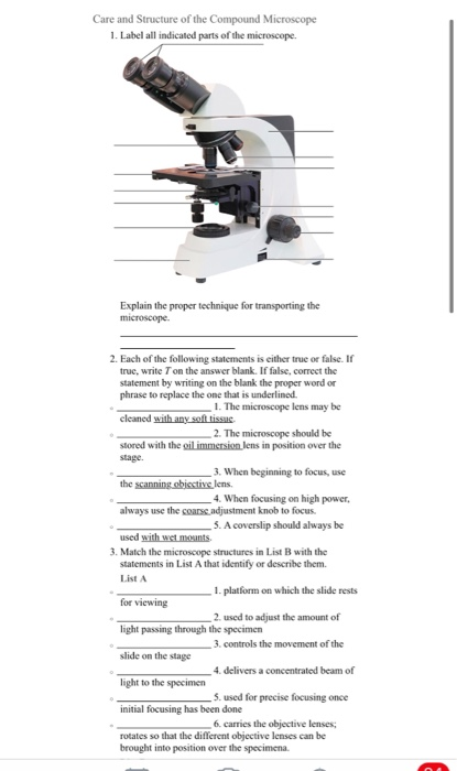
Care and Structure of the Compound Microscope 1. | Chegg.com
Compound Microscope with labels Stock Vector | Adobe Stock Download Compound Microscope with labels Stock Vector and explore similar vectors at Adobe Stock. Adobe Stock Photos Illustrations Vectors Videos Audio Templates Free Premium Editorial Fonts

Diagrams of Microscope | 101 Diagrams
› pmc › articlesClinical-grade computational pathology using weakly ... The medical image analysis field has seen widespread application of deep learning, showing in some cases that clinical impact can be achieved for diagnostic tasks. Notably, ref. 11 reported dermatologist-level diagnosis of dermoscopy images, while ref. 12 showed ophthalmologist-level performance on optical coherence tomography images.
Post a Comment for "38 images of compound microscope with labels"