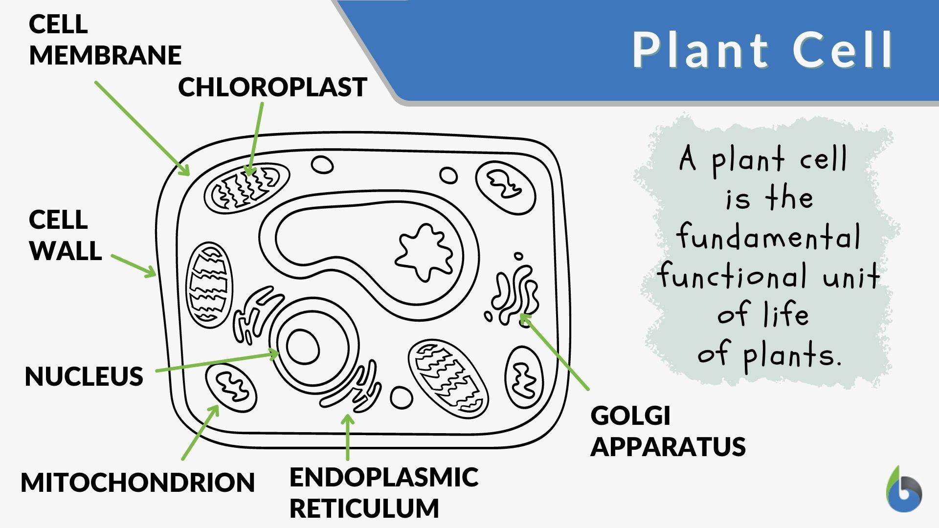
41 diagram of a plant cell without labels
What Are Cyanobacteria, and How Are They Similar or ... - Owlcation Below, we will explore their similarities and differences by comparing their structure and how they perform key functions necessary for life. Figure 1. Labeled diagram of a plant cell. Natalie Lamb 1 / 3 Structure of Plant Cells, Cyanobacteria, and Chloroplasts Cell Type and Structure Plasma Membrane (Cell Membrane) - Genome.gov The plasma membrane, also called the cell membrane, is the membrane found in all cells that separates the interior of the cell from the outside environment. In bacterial and plant cells, a cell wall is attached to the plasma membrane on its outside surface. The plasma membrane consists of a lipid bilayer that is semipermeable.
owlcation.com › stem › 3d-cell-modelHow to Create 3D Plant Cell and Animal Cell Models for ... Sep 10, 2011 · Step 1: Choose Plant Cell vs. Animal Cell. First and foremost, you must decide whether you will create a plant or animal cell. Plant cells and animal cells are shaped differently and contain different parts. The best way to decide? Take a look at some cell diagrams on an interactive site like Cells Alive. This site offers awesome animations of ...

Diagram of a plant cell without labels
DNA explained: Structure, function, and impact on health Virtually every cell in the body contains deoxyribonucleic acid (DNA). It is the genetic code that makes each person unique. DNA carries the instructions for the development, growth, reproduction ... Spatial resolution of an integrated C4+CAM photosynthetic metabolism ... Despite the large number of independent origins of both C 4 and CAM, for the most part, plant lineages tend to evolve one CCM or the other. Over 40 years ago, Portulaca oleracea was identified as the first known C 4 plant that also operates a facultative CAM cycle (C 4 +CAM) in response to drought or changes in photoperiod ().Full integration of C 4 and CAM cycles, whereby both operate in the ... en.wikipedia.org › wiki › History_of_botanyHistory of botany - Wikipedia Progressively more sophisticated scientific technology has aided the development of contemporary botanical offshoots in the plant sciences, ranging from the applied fields of economic botany (notably agriculture, horticulture and forestry), to the detailed examination of the structure and function of plants and their interaction with the ...
Diagram of a plant cell without labels. How to Create Beautiful Diagrams for Your Documents and Presentations Limit the number of colors you use in your diagram as it may make it look chaotic. Instead, stick to 3-4 colors to preserve the readability of the diagram. And you can use different shades of the same color to indicate the relationships among various shapes. Use different colors to provide contrast to different objects. Duodenum: Anatomy, histology, composition, functions | Kenhub Anatomy Sections The duodenum is about 25 to 30 cm long ("twelve fingers' length"), C-shaped and is located in the upper abdomen at the level of L1-L3. The head of the pancreas lies in the C loop. It may be subdivided into four sections: superior part, descending part, horizontal part and ascending part. Cell Cycle - Genome.gov A cell cycle is a series of events that takes place in a cell as it grows and divides. A cell spends most of its time in what is called interphase, and during this time it grows, replicates its chromosomes, and prepares for cell division. The cell then leaves interphase, undergoes mitosis, and completes its division. Diagram of Human Heart and Blood Circulation in It A heart diagram labeled will provide plenty of information about the structure of your heart, including the wall of your heart. The wall of the heart has three different layers, such as the Myocardium, the Epicardium, and the Endocardium. Here's more about these three layers. Epicardium
Vacuole - Genome.gov Definition. 00:00. …. A vacuole is a membrane-bound cell organelle. In animal cells, vacuoles are generally small and help sequester waste products. In plant cells, vacuoles help maintain water balance. Sometimes a single vacuole can take up most of the interior space of the plant cell. › publication › 362082926Plasma membrane-nucleo-cytoplasmic coordination of a receptor ... Jul 01, 2022 · The plant immune system involves cell-surface receptors that detect intercellular pathogen-derived molecules, and intracellular receptors that activate immunity upon detection of pathogen-secreted ... › heart › picture-of-the-heartHuman Heart (Anatomy): Diagram, Function, Chambers, Location ... WebMD's Heart Anatomy Page provides a detailed image of the heart and provides information on heart conditions, tests, and treatments. Eukaryotic Cells Quiz - ProProfs Quiz Eukaryotic cells are known to have a nucleus enclosed within the nuclear membrane, and they form huge and complex organisms. Protozoa, plants, fungi, and animals all have eukaryotic cells. They are classified under the kingdom of Eukaryota. These questions will give you an even better understanding of eukaryotic cells. Go for it! All the best ...
Diatom - Wikipedia Diatom (Neo-Latin diatoma) refers to any member of a large group comprising several genera of algae, specifically microalgae, found in the oceans, waterways and soils of the world.Living diatoms make up a significant portion of the Earth's biomass: they generate about 20 to 50 percent of the oxygen produced on the planet each year, take in over 6.7 billion metric tons of silicon each year from ... Brigitte Zimmer 9Th Grade Easy Plant Cell Diagram Simple Diagram Of A Heart With Labels Label The Picture Of A Human Digestive System Labeled Sketch Of A Chloroplast Internal Female Reproductive System Labelled Diagram Dna Easy Drawing Digestive System Diagram Class 10 Easy Easy Plant Cell And Animal Cell Drawing With Labels Animal Cell Picture With Labeled Parts Histology guide: Definition and slides | Kenhub It is composed of densely packed epithelial cells with only a little extracellular matrix (ECM). The cells are laid down on top of dense irregular connective tissue, the basement membrane (BM). Epithelium is classified by both it's cellular morphology and the number of cell layers. Concept Map Tutorial: How to Create Concept Maps to Visualize Ideas Step 3: Start to Draw the Map. It's recommended to start a concept map from the top and develop it downward, although you can put down your topic at the center and expand it outwards. Either way make sure that the central topic stands out from the rest (use a bigger node, a different color etc.).

a picture of a plant cell with labels | plant cell (diagram & label)(7-2) | Plant Cell ideas ...
Hypodermis of the Skin Anatomy and Physiology - Verywell Health The hypodermis is the innermost layer of the skin. It stores fat and energy, pads and protects the body, attaches skin to the bones and muscle, and is very important in maintaining body temperature. This layer of the skin thins with age, increasing the risk for hypothermia or heat exhaustion. It provides shaping and contouring, and the ...
Plant Cells Vs. Animal Cells (With Diagrams) - Owlcation The most important structures of plant and animal cells are shown in the diagrams below, which provide a clear illustration of how much these cells have in common. The significant differences between plant and animal cells are also shown, and the diagrams are followed by more in-depth information. Diagram of an animal cell Doc Sonic
Pancreas histology: Exocrine & endocrine parts, function | Kenhub Pancreatic Islets are spherical clusters of polygonal endocrine cells. On a pancreas histological slide stained with H&E, they appear as large, pale-staining cells enveloped by intensely staining, basophilic pancreatic acini. The cells of the islets are connected to each other with desmosomes and gap junctions, forming bands or cords of cells.
photosynthesis | Definition, Formula, Process, Diagram, Reactants ... In chemical terms, photosynthesis is a light-energized oxidation-reduction process. (Oxidation refers to the removal of electrons from a molecule; reduction refers to the gain of electrons by a molecule.) In plant photosynthesis, the energy of light is used to drive the oxidation of water (H 2 O), producing oxygen gas (O 2 ), hydrogen ions (H ...
› standard-level › topic-1-cellProkaryotic Cells | BioNinja Cell membrane – Semi-permeable and selective barrier surrounding the cell Cell wall – rigid outer covering made of peptidoglycan; maintains shape and prevents bursting (lysis) Slime capsule – a thick polysaccharide layer used for protection against dessication (drying out) and phagocytosis
NCERT Book Class 8 Science Chapter 8 Cell - Structure and Functions January 7, 2022. in 8th Class. Reading Time: 3 mins read. 0. NCERT Book for Class 8 Science Chapter 8 Cell - Structure and Functions is available for reading or download on this page. Students who are in Class 8 or preparing for any exam which is based on Class 8 Science can refer NCERT Science Book for their preparation.
Label The Cell Diagram Plant - vrl.serviziocatering.trieste.it plant cells: they have one or more, comparatively very smaller vacuoles label a diagram of an animal cell and a plant cell; a diagram showing how proteins are produced by ribosomes, and finally packaged by the golgi this worksheet helps students learn the parts of the cell it includes a diagram of an animal cell and a plant cell for labeling for …

Plant Cell Science Diagram Clipart Set 300 dpi School | Etsy | Plant cell diagram, Science ...
What is a Dichotomous Key | Step-by-Step Guide with Editable ... - Creately Step 5: Draw a dichotomous key diagram. You can either create a text-based dichotomous key or a graphical one where you can even use images of the specimen you are trying to identify. Here you can use a tree diagram or a flowchart as in the examples below. Step 6: Test it out . Once you have completed your dichotomous key, test it out to see if ...
Different Size, Shape and Arrangement of Bacterial Cells In shape they may principally be Rods (bacilli), Spheres (cocci), and Spirals (spirillum). Size of Bacterial Cell The average diameter of spherical bacteria is 0.5-2.0 µm. For rod-shaped or filamentous bacteria, length is 1-10 µm and diameter is 0.25-1 .0 µm. E. coli , a bacillus of about average size is 1.1 to 1.5 µm wide by 2.0 to 6.0 µm long.

Plant Cell Science Diagram Clipart Set 300 dpi School | Etsy | Science diagrams, Plant cell ...
Flowering plant - Wikipedia Cross-section of a stem of the angiosperm flax: 1. pith, 2. protoxylem, 3. xylem, 4. phloem, 5. sclerenchyma ( bast fibre ), 6. cortex, 7. epidermis Angiosperm stems are made up of seven layers as shown on the right. The amount and complexity of tissue-formation in flowering plants exceeds that of gymnosperms.

Cell Wall Found In Plant Animal Or Both - Both Plant And Animal Cell Wall Java Virtual Machine ...
onlinelibrary.wiley.com › doi › 10Single‐cell transcriptome atlas reveals developmental ... Jul 10, 2022 · Single cell captured from 1 L and 3 L were processed using 10× Genomics Chromium Single Cell 3′ Solution. The Cell Ranger output was loaded into Seurat (version 3.1.1) which was used for dimensional reduction, clustering and analysis of scRNA-seq data. Overall, cells passed the quality control threshold when the following criteria were met.
plant cell diagram : text, images, music, video | Glogster EDU - Interactive multimedia posters
Plant Tissues: Name, Types, Functions, Diagram - Embibe On the basis of position in the plant body, meristematic tissue is divided into the following types: a. Apical Meristem 1. This meristem is located at the growing tips of main and lateral roots and shoots. These cells are responsible for the linear growth of an organ. 2. They are mostly primary meristems. 3. E.g., Shoot apex and root apex. b.
Introduction to Scale Insects | University of Maryland Extension - UMD Key Points. Considered pests, scale are sucking insects that consume sap or plant cell contents. They are categorized as either armored (hard) scale or soft scale, and this distinction determines the damage they can cause and how they are best controlled.; Low populations tend to go undetected. High populations can cause plant damage, such as leaf yellowing, plant stunting, or branch dieback.
Cell Diagram Not Labeled - biology is amazing cell structures, amoeba ... Cell Diagram Not Labeled - 16 images - cell diagram, a typical cell labeled royalty free stock photos image 26503298, chapter 3 page 4 histologyolm, cytokinesis in animal cells illustration stock image c023 8854,
WHMIS 2015 - Pictograms : OSH Answers - Canadian Centre for ... What is a pictogram? Pictograms are graphic images that immediately show the user of a hazardous product what type of hazard is present. With a quick glance, you can see, for example, that the product is flammable, or if it might be a health hazard. Most pictograms have a distinctive red "square set on one of its points" border.
Structure and parts of a sperm cell - inviTRA Structure and parts of a sperm cell 0 This labelled diagram shows the structure of a sperm cellin detail, which has the following parts: Head With its spheric shape, it consists of a large nucleus, which at the same time contains an acrosome. The nucleus contains the genetic information and 23 chromosomes.

Biology Pictures: Plant Cell Diagram | Homeschool Helps | Pinterest | Marine biology, Pictures ...
DeLTa-Seq: direct-lysate targeted RNA-Seq from crude tissue lysate ... Reducing regents inhibit RNA degradation in plant, yeast, and animal lysates. For direct-lysate RT from plant tissues, we trialed three reagents [CL buffer [], CellAmp (TaKaRa, Kusatsu, Japan), and SuperPrep (TOYOBO, Osaka, Japan)] designed for direct-lysate RT from cultured mammalian cells.Arabidopsis thaliana seedlings were homogenized in these reagents, followed by a 1 h incubation at 22 °C.


Post a Comment for "41 diagram of a plant cell without labels"